TUSC3
| TUSC3 | ||||||||||||||||||||||||||||||||||||||||||||||||||||||
|---|---|---|---|---|---|---|---|---|---|---|---|---|---|---|---|---|---|---|---|---|---|---|---|---|---|---|---|---|---|---|---|---|---|---|---|---|---|---|---|---|---|---|---|---|---|---|---|---|---|---|---|---|---|---|
 | ||||||||||||||||||||||||||||||||||||||||||||||||||||||
| Identifiers | ||||||||||||||||||||||||||||||||||||||||||||||||||||||
| Aliases | TUSC3, D8S1992, M33, MRT22, MRT7, N33, OST3A, tumor suppressor candidate 3, MagT2, SLC58A2 | |||||||||||||||||||||||||||||||||||||||||||||||||||||
| External IDs | OMIM: 601385; MGI: 1933134; GeneCards: TUSC3 | |||||||||||||||||||||||||||||||||||||||||||||||||||||
| ||||||||||||||||||||||||||||||||||||||||||||||||||||||
| ||||||||||||||||||||||||||||||||||||||||||||||||||||||
| ||||||||||||||||||||||||||||||||||||||||||||||||||||||
| ||||||||||||||||||||||||||||||||||||||||||||||||||||||
| ||||||||||||||||||||||||||||||||||||||||||||||||||||||
| ||||||||||||||||||||||||||||||||||||||||||||||||||||||
| Wikidata | ||||||||||||||||||||||||||||||||||||||||||||||||||||||
| ||||||||||||||||||||||||||||||||||||||||||||||||||||||


Tumor suppressor candidate 3 (TUSC3), formerly known as N33 or OST3A, is a protein that in humans is encoded by the TUSC3 gene on chromosome 8p22.[5]
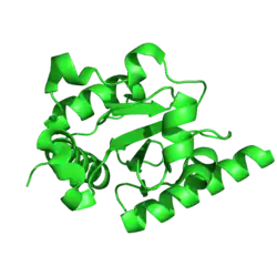
Structurally, TUSC3 is an integral membrane protein localized to the endoplasmic reticulum (ER) that contains an N-terminal thioredoxin-like domain.[6]
Functionally, TUSC3 exhibits a dual biochemical role. It acts as an accessory unit of the oligosaccharyltransferase (OST) complex, facilitating N-linked glycosylation of nascent proteins.[6] In addition, TUSC3 forms a complex with the ER magnesium transporter ERMA(TMEM94) and is required for efficient magnesium uptake into the ER lumen. Disruption of this TUSC3--ERMA complex leads to magnesium depletion, activation of the PERK--eIF2α stress pathway, impaired synaptic protein translation and neurodevelopmental deficits, linking TUSC3 loss to autosomal recessive intellectual disability (ARID).[7]
TUSC3 has also been studied as a candidate tumor suppressor gene, with loss or silencing associated with progression in several human cancers.[8][9][10]
Structure
TUSC3 is a multi-pass transmembrane protein of the ER. It consists of a large N-terminal domain that faces the ER lumen and a C-terminal region containing four transmembrane helices that anchor the protein in the membrane. The luminal domain adopts a thioredoxin-like fold and contains a conserved redox-active Cys-X-X-Cys motif. The C-terminal transmembrane bundle allows TUSC3 to be incorporated into the STT3B-containing OST complex as one of two alternative accessory subunits (the other being MAGT1).[6][11]
Function
Role in N-linked glycosylation
N-linked glycosylation is the process in which a preassembled oligosaccharide (sugar chain) is attached to the nitrogen atom of specific asparagine residues within Asn-X-Ser/Thr sequons of nascent polypeptides. This modification occurs in the ER and is catalyzed by the OST complex. Disruptions to this pathway cause congenital disorders of glycosylation (CDG).[12][13]
Molecular mechanism
Mammalian cells express two distinct forms of the OST complex: STT3A, which acts co-translationally at the translocon, and STT3B, which acts post-translationally to glycosylate acceptor sites skipped by STT3A.[14] TUSC3, along with its paralog MAGT1, specifically associates with the STT3B complex.[14]
Substrate specificity and redundancy
TUSC3 and MAGT1 share high sequence identity and can act redundantly within the STT3B-containing complex.[15] Simultaneous loss of both TUSC3 and MAGT1 severely compromises STT3B-dependent glycosylation, leading to increased protein misfolding in the ER lumen, activation of the unfolded protein response (UPR), and ER stress-induced apoptosis.[14][8]
Knockout of TUSC3 alone does not cause systemic N-glycan loss or alter serum glycoprotein patterns in most tissues, because MAGT1 can compensate for it’s absence.[8] Although TUSC3 and MAGT1 show functional redundancy in vitro, their endogenous expression patterns differ: MAGT1 is predominantly expressed in immune cells, whereas TUSC3 is highly enriched in the central nervous system.[15] As a result, loss of TUSC3 cannot be adequately compensated for in brain tissue, leading to neurological pathology.[15]
Discovery and initial classification as a congenital disorder of glycosylation (CDG)
The first disease association for TUSC3 came through its role in glycosylation. Because TUSC3 encodes a non-catalytic subunit of the ER-bound OST complex, two research groups that independently identified TUSC3 mutations in patients with intellectual disability (ID) in 2008 proposed that the condition represented a CDG. This classification, sometimes referred to as TUSC3-CDG, was based on TUSC3’s position within the glycosylation machinery rather than direct biochemical evidence of a glycosylation defect in patients.[13][16]
At the time, this inference appeared reasonable: most CDGs are identified through abnormal glycosylation patterns detectable on routine clinical testing (such as isoelectric focusing of serum transferrin), and several other CDGs cause neurodevelopmental impairment through disrupted glycosylation of brain proteins. Researchers therefore expected TUSC3 loss to behave similarly. However, both founding studies reported normal results on standard glycosylation screens in affected patients—an atypical finding for a CDG that left the actual disease mechanism unresolved for more than a decade.[13][16]
Role in ER magnesium homeostasis
(in progress)
Historical identification as a Mg2+ transporter
(in progress)
Regulation of ERMA(TMEM94)
(in progress)
Functional consequences
(in progress)
Clinical significance
Intellectual disability
(in progress)
Genetics
(in progress)
Clinical phenotype
(in progress)
Mechanistic link
(in progress)
Role in Cancer
A candidate tumor suppressor gene. TUSC3 is located within a deleted region of a metastatic prostate cancer. The gene is expressed in most nonlymphoid human tissues including prostate, lung, liver, and colon. Expression was also detected in many epithelial tumor cell lines. Two transcript variants encoding distinct isoforms have been identified for this gene.[17]
References
- 1 2 3 GRCh38: Ensembl release 89: ENSG00000104723 – Ensembl, May 2017
- 1 2 3 GRCm38: Ensembl release 89: ENSMUSG00000118664 – Ensembl, May 2017
- ↑ "Human PubMed Reference:". National Center for Biotechnology Information, U.S. National Library of Medicine.
- ↑ "Mouse PubMed Reference:". National Center for Biotechnology Information, U.S. National Library of Medicine.
- ↑ "TUSC3 tumor suppressor candidate 3 [Homo sapiens (human)] - Gene - NCBI". www.ncbi.nlm.nih.gov. Retrieved 2026-08-25.
- 1 2 3 Mohorko E, Owen RL, Malojčić G, Brozzo MS, Aebi M, Glockshuber R (April 2014). "Structural basis of substrate specificity of human oligosaccharyl transferase subunit N33/Tusc3 and its role in regulating protein N-glycosylation". Structure. 22 (4). London: 590–601. doi:10.1016/j.str.2014.02.013. PMID 24685145.
- ↑ Park G, Kim N, Kim SY, Lee H, Li CM, Seol JH, et al. (November 2025). "TUSC3 regulates ERMA-mediated Mg2+ uptake for synaptic function and neurodevelopment". Nature Communications. 16 (1) 9752. doi:10.1038/s41467-025-65668-1. PMC 12594762. PMID 41203647.
- 1 2 3 Vašíčková K, Horak P, Vaňhara P (March 2018). "TUSC3: functional duality of a cancer gene". Cellular and Molecular Life Sciences. 75 (5): 849–857. doi:10.1007/s00018-017-2660-4. PMC 11105401. PMID 28929175.
- ↑ Kratochvílová K, Horak P, Ešner M, Souček K, Pils D, Anees M, et al. (September 2015). "Tumor suppressor candidate 3 (TUSC3) prevents the epithelial-to-mesenchymal transition and inhibits tumor growth by modulating the endoplasmic reticulum stress response in ovarian cancer cells". International Journal of Cancer. 137 (6): 1330–1340. doi:10.1002/ijc.29502. PMID 25735931.
- ↑ Yu X, Zhai C, Fan Y, Zhang J, Liang N, Liu F, et al. (September 2017). "TUSC3: a novel tumour suppressor gene and its functional implications". Journal of Cellular and Molecular Medicine. 21 (9): 1711–1718. doi:10.1111/jcmm.13128. PMC 5571513. PMID 28272772.
- ↑ "UniProt". UniProt. Retrieved 2026-08-27.
- ↑ Mohorko J, Glockshuber R, Aebi M (October 2011). "Oligosaccharyltransferase subunit MAGT1 interacts with STT3B to promote N-glycosylation". Journal of Biological Chemistry. 286 (42): 36162–36170. doi:10.1074/jbc.M111.283838 – via JBC.
- 1 2 3 Molinari F, Foulquier F, Tarpey PS, Morelle W, Boissel S, Teague J, et al. (May 2008). "Oligosaccharyltransferase-subunit mutations in nonsyndromic mental retardation". American Journal of Human Genetics. 82 (5): 1150–1157. doi:10.1016/j.ajhg.2008.03.021. PMC 2427205. PMID 18455129.
- 1 2 3 Cherepanova NA, Shrimal S, Gilmore R (August 2014). "Oxidoreductase activity is necessary for N-glycosylation of cysteine-proximal acceptor sites in glycoproteins". The Journal of Cell Biology. 206 (4): 525–539. doi:10.1083/jcb.201404085. PMC 4137057. PMID 25135935.
- 1 2 3 Blommaert E, Péanne R, Cherepanova NA, Rymen D, Staels F, Jaeken J, et al. (May 2019). "Mutations in MAGT1 lead to a glycosylation disorder with a variable phenotype". Proceedings of the National Academy of Sciences of the United States of America. 116 (20): 9865–9870. doi:10.1073/pnas.1817815116. PMC 6525510. PMID 31036665.
- 1 2 Garshasbi M, Hadavi V, Habibi H, Kahrizi K, Kariminejad R, Behjati F, et al. (May 2008). "A Defect in the TUSC3 Gene Is Associated with Autosomal Recessive Mental Retardation". The American Journal of Human Genetics. 82 (5): 1158–1164. doi:10.1016/j.ajhg.2008.03.018. PMC 2651624. PMID 18452889 – via PubMed.
- ↑ "Entrez Gene: TUSC3 tumor suppressor candidate 3".
Further reading
- Pak BJ, Park H, Chang ER, Pang SC, Graham CH (September 1998). "Differential display analysis of oxygen-mediated changes in gene expression in first trimester human trophoblast cells". Placenta. 19 (7): 483–488. doi:10.1016/S0143-4004(98)91041-4. PMID 9778121.
- Kelleher DJ, Karaoglu D, Mandon EC, Gilmore R (2003). "Oligosaccharyltransferase isoforms that contain different catalytic STT3 subunits have distinct enzymatic properties". Molecular Cell. 12 (1): 101–111. doi:10.1016/S1097-2765(03)00243-0. PMID 12887896.
- Anderson NL, Polanski M, Pieper R, Gatlin T, Tirumalai RS, Conrads TP, et al. (April 2004). "The human plasma proteome: a nonredundant list developed by combination of four separate sources". Molecular & Cellular Proteomics. 3 (4): 311–326. doi:10.1074/mcp.M300127-MCP200. PMID 14718574.
- Colland F, Jacq X, Trouplin V, Mougin C, Groizeleau C, Hamburger A, et al. (July 2004). "Functional proteomics mapping of a human signaling pathway". Genome Research. 14 (7): 1324–1332. doi:10.1101/gr.2334104. PMC 442148. PMID 15231748.
- Shibatani T, David LL, McCormack AL, Frueh K, Skach WR (April 2005). "Proteomic analysis of mammalian oligosaccharyltransferase reveals multiple subcomplexes that contain Sec61, TRAP, and two potential new subunits". Biochemistry. 44 (16): 5982–5992. doi:10.1021/bi047328f. PMID 15835887.
- Rual JF, Venkatesan K, Hao T, Hirozane-Kishikawa T, Dricot A, Li N, et al. (October 2005). "Towards a proteome-scale map of the human protein-protein interaction network". Nature. 437 (7062): 1173–1178. Bibcode:2005Natur.437.1173R. doi:10.1038/nature04209. PMID 16189514. S2CID 4427026.
- Pils D, Horak P, Gleiss A, Sax C, Fabjani G, Moebus VJ, et al. (December 2005). "Five genes from chromosomal band 8p22 are significantly down-regulated in ovarian carcinoma: N33 and EFA6R have a potential impact on overall survival". Cancer. 104 (11): 2417–2429. doi:10.1002/cncr.21538. PMID 16270321. S2CID 26030376.
- Guervós MA, Marcos CA, Hermsen M, Nuño AS, Suárez C, Llorente JL (2007). "Deletions of N33, STK11 and TP53 are involved in the development of lymph node metastasis in larynx and pharynx carcinomas". Cellular Oncology. 29 (4): 327–334. doi:10.1155/2007/635962. PMC 4618212. PMID 17641416.