PAAT (protein)
| C10orf88 | ||||||||||||||||||||||||||||||||||||||||||||||||||||||
|---|---|---|---|---|---|---|---|---|---|---|---|---|---|---|---|---|---|---|---|---|---|---|---|---|---|---|---|---|---|---|---|---|---|---|---|---|---|---|---|---|---|---|---|---|---|---|---|---|---|---|---|---|---|---|
| Identifiers | ||||||||||||||||||||||||||||||||||||||||||||||||||||||
| Aliases | C10orf88, chromosome 10 open reading frame 88, PAAT | |||||||||||||||||||||||||||||||||||||||||||||||||||||
| External IDs | MGI: 1915527; GeneCards: C10orf88 | |||||||||||||||||||||||||||||||||||||||||||||||||||||
| ||||||||||||||||||||||||||||||||||||||||||||||||||||||
| ||||||||||||||||||||||||||||||||||||||||||||||||||||||
| ||||||||||||||||||||||||||||||||||||||||||||||||||||||
| ||||||||||||||||||||||||||||||||||||||||||||||||||||||
| Wikidata | ||||||||||||||||||||||||||||||||||||||||||||||||||||||
| ||||||||||||||||||||||||||||||||||||||||||||||||||||||


PAAT (protein associated with ABC transporters), also known as C10orf88 (chromosome 10 open reading frame 88), is a human protein encoded by the gene of the same name.[5] It contains an ABC transporter domain predicted to associate with Type II ABC transporters.[6] PAAT physically interacts with mitochondrial inner membrane ABC transporters and has a demonstrated role in ferric nutrient transport and heme biosynthesis. It is primarily expressed in the cerebral cortex, testis, and thymus, and localizes to the cytoplasm and nucleus. The gene has been associated with a rare form of Alzheimer's disease.
Evolution
The earliest detectable ortholog of PAAT occurs in the subphylum Cephalochordata, specifically the European lancelet (Branchiostoma lanceolatum).[7] This organism represents one of the most basal chordates, diverging from the vertebrate lineage approximately 588 million years ago (MYA), likely during the Cambrian period, though some phylogenetic models place this divergence as late as the Cretaceous. The lancelet ortholog shares only 21% sequence identity and 38% similarity with the human protein, with a corrected sequence divergence (CSD) of 156%. A comparable pattern holds in the Brook lamprey (Lampetra planeri), another early-branching chordate.[8] No orthologs have been identified in invertebrates or prokaryotes, therefore PAAT appears to have a chordate-specific origin. PAAT is also absent in Sarcopterygii, despite being present in Actinopterygii; these groups differ in the presence of limb buds supported by articulated appendicular skeletons.[9]

Sequence identity increases progressively with greater relatedness, with mammals averaging approximately 80% identity and the lowest overall CSD values. This pattern is consistent with purifying selection in lineages reliant on hemoglobin-based oxygen transport, given PAAT's proposed role in heme transport.[10]
Compared to model proteins with well-characterized evolutionary rates, Cytochrome C (slow) and Fibrinogen alpha (fast), PAAT tracks closer to the fast-evolving end, with CSD values rising steeply with phylogenetic distance. This rapid divergence may reflect substantial functional variation across taxa, particularly given that heme transport roles appear prominent only in humans.

| Class | Seq | Genus species | Common name | Taxon | Date of Div (MYA) | CorSeqDiv% | Accession # | Seq Length (aa) | Seq ID (%) | Seq Sim (%) |
|---|---|---|---|---|---|---|---|---|---|---|
| Mammalia | Hsa | Homo sapiens | Human | Primates | 0 | 0 | NP_079218 | 445 | 100 | 100 |
| Mammalia | Mmu | Mus musculus | House mouse | Rodentia | 87 | 34.24903089 | NP_080931.1 | 444 | 71 | 83 |
| Mammalia | Egl | Eubalaena glacialis | North Atlantic right whale | Artiodactyl | 94 | 22.31435513 | XP_061048496.1 | 445 | 80 | 88 |
| Mammalia | Fca | Felis catus | House cat | Carnivora | 94 | 28.76820725 | XP_038614534.1 | 444 | 75 | 85 |
| Reptilia | Eor | Emys orbicularis | European pond turtle | Testudines | 319 | 65.39264674 | XP_065264191.1 | 462 | 52 | 68 |
| Reptilia | Pra | Podarcis raffonei | Aeolian wall lizard | Lacertidae | 319 | 69.31471806 | XP_053244946.1 | 456 | 47 | 59 |
| Reptilia | Pca | Pituophis catenifer | Gopher snake | Squamata | 319 | 84.39700703 | XP_070801900.1 | 454 | 43 | 59 |
| Aves | Gca | Gymnogyps californianus | Californian Condor | Accipitriformes | 319 | 75.50225843 | XP_050754500.1 | 445 | 47 | 62 |
| Aves | Aph | Agelaius phoeniceus | Red-winged blackbird | Passeriformes | 319 | 82.09805521 | XP_054493097.1 | 442 | 44 | 60 |
| Aves | Sca | Struthio camelus | Common ostrich | Struthioniformes | 319 | 84.39700703 | XP_068807255.1 | 481 | 43 | 59 |
| Amphibia | Ame | Ambystoma mexicanum | Axolotl | Urodela | 352 | 107.8809661 | XP_069468114.1 | 405 | 34 | 52 |
| Amphibia | Hya | Hyla sarda | Tyrrhenian tree frog | Anura | 352 | 110.8662625 | XP_056385755.1 | 409 | 33 | 50 |
| Amphibia | Gse | Geotrypetes seraphini | Gaboon caecilian | Gymnophiona | 352 | 117.1182982 | XP_033799666.1 | 371 | 31 | 45 |
| Actinopterygii | Psp | Polyodon spathula | American paddlefish | Acipenseriformes | 429 | 113.9434283 | XP_041125204.1 | 433 | 32 | 51 |
| Actinopterygii | Eca | Erpetoichthys calabaricus | Reedfish | Polypteriformes | 429 | 146.967597 | XP_028651065.1 | 438 | 23 | 45 |
| Chondrichthyes | Sti | Stegostoma tigrinum | Zebra shark | Orectolobiformes | 462 | 104.9822124 | XP_048407415.1 | 439 | 35 | 52 |
| Chondrichthyes | Hfr | Heterodontus francisci | Horn shark | Heterodontiformes | 462 | 104.9822124 | XP_067908923.1 | 441 | 35 | 50 |
| Chondrichthyes | Pja | Pristiophorus japonicus | Japanese sawshark | Pristiophoriformes | 462 | 107.8809661 | XP_070732813.1 | 448 | 34 | 51 |
| Petromyzontida | Lpl | Lampetra planeri | Brook lamprey | Petromyzontiformes | 563 | 156.0647748 | CAL9842239 | 499 | 21 | 35 |
| Leptocardii | Bla | Branchiostoma lanceolatum | European lancelet | Branchiostomatidae | 588 | 156.0647748 | CAH1258791.1 | 414 | 21 | 38 |
Gene
PAAT maps to chromosome 10 at cytogenetic locus 10q26.13. The gene spans 2,913 base pairs, comprises six exons and five introns, and encodes a 445-amino acid protein. No isoforms or splice variants have been identified. The mature mRNA is 1,497 nucleotides after splicing.
Transcription Factors
The PAAT promoter contains binding sites for transcription factors associated with neural, immune, and reproductive contexts, consistent with the gene's expression profile. Key factors include NRF1, NR2C2, ELK4, ETV4, TFAP2A, and IRF3.
Protein
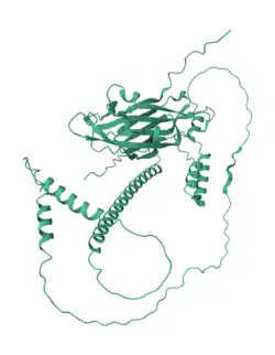
PAAT functions as a novel ATPase, catalyzing ATP hydrolysis to regulate iron homeostasis. It contains two domains: a primary PAAT-like functional domain, and a secondary domain shared with other ABC transporters (designated "ABC Transport 1"). The ABC Transport 1 domain is predominantly beta sheet in secondary structure, while the C-terminus is largely alpha-helical. The protein has a theoretical isoelectric point of 6.0 and a predicted molecular weight of 49.2 kDa.[11] No isoforms, disulfide bonds, or oligomerization have been identified.

Subcellular Localization
PSORT II[12] and DeepLoc[13] both predict mainly nuclear localization (~60% confidence). Their secondary predictions branch: PSORT II favors mitochondrial (17.4%) and cytoplasmic (13%) signals, while DeepLoc assigns a stronger cytoplasmic probability (41%). No mitochondrial targeting sequences or cleavage sites have been detected, which weighs against exclusive mitochondrial localization. WoLF PSORT[14] confirms nuclear and cytoplasmic localization as the two primary compartments. Analysis of known interacting proteins similarly clusters in the nucleus and cytoplasm, with mitochondria as a secondary possibility. These predictions are consistent across orthologs.
Protein Interactions
PAAT is experimentally confirmed to interact with the mitochondrial inner membrane transporters ABCB7, ABCB8, and ABCB10, but not with ABCB1 or ABCG2.[15] The STRING database identifies ten interacting proteins, five with experimental support: CLTC, GDI2, CCDC91, NCKAP1, and LILRB4.[16] Most interactions are supported by co-expression data, except TEX9, NADSYN1, and LILRB4; all but POLR3F are text-mining supported. CLTC shows the most consistent co-expression across GEO microarray datasets.[17] NADSYN1 and CYP2R1 may interact with PAAT through the vitamin D pathway.[18]
Post-Translational Modifications
PAAT is predicted to undergo phosphorylation at multiple serine residues (S11, S150, S201),[19] lysine acetylation at K25,[20] and both N- and O-linked glycosylation.[21] Degron motifs are present for both SCF-βTrCP1 and APC/C complexes.[22] Functional motifs include 14-3-3 and SH2 binding sites, a PP1 phosphatase docking site,[23] and a nuclear export signal.[24] No signal peptides, lipid anchors, or organelle-targeting sequences have been detected.
Expression
Gene Expression
PAAT is expressed across a broad range of human tissues,[25] with elevated RNA levels in the testis (RPKM: 4.006), brain (RPKM: 1.916), and thymus per Human Protein Atlas RNA-seq data.[26]
GEO datasets (GDS1096, GDS596, GDS3113) refine this picture further. GDS1096 shows broad brain expression with peaks in the thalamus and amygdala, alongside strong testis expression. GDS596 highlights olivary ganglia, superior cervical ganglia, and dorsal root ganglia. GDS3113 shows high expression in both the fetal brain and the testis. Total RNA-seq across 20 human tissues confirms significant expression in the cerebellum and thymus.[27]
During fetal development, circular RNA data indicate early-stage PAAT expression across tissues without marked tissue specificity, in which a broad developmental role has been proposed. [28]
Expression Quantitative Trait Loci (eQTLs)
Several eQTLs regulate PAAT expression in a tissue-dependent manner. The variant chr10_123305622_C_A_b38 has a strong negative normalized effect score (NES: −1.0) in EBV-transformed lymphocytes, while chr10_123424873_AT_A_b38 shows a positive NES (0.86) in the esophagus muscularis. Brain-region variants, including those in the caudate and cerebellar hemisphere, exert negative regulatory effects. A testis-specific positive eQTL (NES: 0.39) is also present. Some eQTL-affected tissue, such as EBV-transformed lymphocytes and esophageal tissue, is not well represented at the protein level, which may reflect disease- or context-specific regulatory activity.
Protein Expression
Immunohistochemical data show cytoplasmic staining in the testis and pancreas. Testicular expression is confined to subsets of cells within the seminiferous ducts; pancreatic expression is prominent in exocrine glandular cells. PaxDb[29] places PAAT protein abundance near the proteome average, with higher levels in the brain, testis, lymph nodes, and ovaries, which are broadly consistent with the RNA expression data.
Condition-Specific Expression
PAAT expression changes in several diseases and differentiation contexts. It is upregulated in redifferentiated podocytes derived from amniotic kidney progenitor cells, a finding that may indicate a role in renal cell maturation.[30] In skeletal muscle from individuals with Myotonic Dystrophy Type 2 (DM2), PAAT is downregulated.[31] It is also upregulated in peripheral blood mononuclear cells during Simian Immunodeficiency Virus (SIV) infection, particularly following PD-1 antibody treatment.[32]
Function
Yang et al. (2014) characterized PAAT as a trans-regulator of mitochondrial ABC transporters with intrinsic ATPase activity.[15] The protein localizes to both the cytoplasm and mitochondria and selectively interacts with ABCB7, ABCB8, and ABCB10, but not ABCB1 or ABCG2, to modulate ferric nutrient transport and heme biosynthesis. Through ATP hydrolysis, PAAT maintains mitochondrial membrane potential and iron homeostasis; its depletion increases oxidative DNA damage and triggers cell death.
Clinical Significance
Alzheimer's Disease
PAAT has been linked to a rare familial form of Alzheimer's disease designated AD15, which presents with amyloid-β plaques but lacks neurofibrillary tangles.[33]
Vitamin D Metabolism
PAAT has been proposed to interact with Cytochrome P450 during hydroxylation in the Vitamin D cycle,[34] with dysregulation associated with Vitamin D deficiency.[35]
Lipid Metabolism
GWAS data associate PAAT variants rs7904973 and rs12246352 with lower LDL and Apolipoprotein B levels in humans.[36][37] In rat models, PAAT has been linked to 2-Methylbutyryl-CoA Dehydrogenase Deficiency (a BCAA catabolism disorder) and craniosynostosis.
References
- 1 2 3 GRCh38: Ensembl release 89: ENSG00000119965 – Ensembl, May 2017
- 1 2 3 GRCm38: Ensembl release 89: ENSMUSG00000040177 – Ensembl, May 2017
- ↑ "Human PubMed Reference:". National Center for Biotechnology Information, U.S. National Library of Medicine.
- ↑ "Mouse PubMed Reference:". National Center for Biotechnology Information, U.S. National Library of Medicine.
- ↑ "C10orf88 chromosome 10 open reading frame 88 [Homo sapiens (Human)] – Gene – NCBI".
- ↑ Locher, K. P. (2008). "Structure and mechanism of ATP-binding cassette transporters". Philosophical Transactions of the Royal Society of London. Series B, Biological Sciences. 364 (1514): 239–245. doi:10.1098/rstb.2008.0125. PMC 2674090. PMID 18957379.
- ↑ "BLAST: Basic Local Alignment Search Tool".
- ↑ "BLAST: Basic Local Alignment Search Tool".
- ↑ Freitas, Renata; Zhang, Guangjun; Cohn, Martin J. (2006). "Evidence that mechanisms of fin development evolved in the midline of early vertebrates". Nature. 442 (7106): 1033–7. Bibcode:2006Natur.442.1033F. doi:10.1038/nature04984. PMID 16878142.
- ↑ Severance, Scott; Hamza, Iqbal (2009). "Trafficking of Heme and Porphyrins in Metazoa". Chemical Reviews. 109 (10): 4596–4616. doi:10.1021/cr9001116. PMC 2769250. PMID 19764719.
- ↑ "Expasy – Compute pI/MW".
- ↑ "PSORT II server – GenScript".
- ↑ "688BA73A0025637599B4014A expired".
- ↑ "AETb6ef5272c9c7d097714e6583e1d3973c.cleaned.fasta PSORT verbose output".
- 1 2 Yang, X.; Yang, J.; Li, L.; Sun, L.; Yi, X.; Han, X.; Si, W.; Yan, R.; Chen, Z.; Xie, G.; Li, W.; Shang, Y.; Liang, J. (2014). "PAAT, a novel ATPase and trans-regulator of mitochondrial ABC transporters, is critically involved in the maintenance of mitochondrial homeostasis". FASEB Journal. 28 (11): 4821–34. doi:10.1096/fj.14-254045. PMID 25063848.
- ↑ "C10orf88 protein (Human) – STRING interaction network".
- ↑ Huttlin EL, Bruckner RJ, Navarrete-Perea J, Cannon JR, Baltier K, Gebreab F, Gygi MP, Thornock A, Zarraga G, Tam S, Szpyt J, Gassaway BM, Panov A, Parzen H, Fu S, Golbazi A, Maenpaa E, Stricker K, Guha Thakurta S, Zhang T, Rad R, Pan J, Nusinow DP, Paulo JA, Schweppe DK, Vaites LP, Harper JW, Gygi SP (May 2021). "Dual proteome-scale networks reveal cell-specific remodeling of the human interactome". Cell. 184 (11): 3022–40. doi:10.1016/j.cell.2021.04.011. PMC 8165030. PMID 33961781.
- ↑ Ahn J, Yu K, Stolzenberg-Solomon R, Simon KC, McCullough ML, Gallicchio L, Jacobs EJ, Ascherio A, Helzlsouer K, Jacobs KB, Li Q, Weinstein SJ, Purdue M, Virtamo J, Horst R, Wheeler W, Chanock S, Hunter DJ, Hayes RB, Kraft P, Albanes D (July 2010). "Genome-wide association study of circulating vitamin D levels". Hum Mol Genet. 19 (13): 2739–45. doi:10.1093/hmg/ddq155. PMC 2883344. PMID 20418485.
- ↑ "C10orf88 (Human)".
- ↑ http://www.pail.biocuckoo.org/
- ↑ "688BA8F500256697E571B59F expired".
- ↑ "Degronopedia".
- ↑ "Elm – Q9H8K7".
- ↑ "NetNES 1.1 – DTU Health Tech – Bioinformatic Services".
- ↑ "Search results < Expression Atlas < EMBL-EBI".
- ↑ "C10orf88 Gene Expression – Gene – NCBI".
- ↑ "C10orf88 Gene Expression – Gene – NCBI".
- ↑ "C10orf88 Gene Expression - Gene - NCBI".
- ↑ "PaxDb: Protein Abundance Database".
- ↑ Da Sacco, Stefano; Lemley, Kevin V.; Sedrakyan, Sargis; Zanusso, Ilenia; Petrosyan, Astgik; Peti-Peterdi, Janos; Burford, James; De Filippo, Roger E.; Perin, Laura (2013). "A Novel Source of Cultured Podocytes". PLOS ONE. 8 (12) e81812. Bibcode:2013PLoSO...881812D. doi:10.1371/journal.pone.0081812. PMC 3861313. PMID 24349133.
- ↑ Screen, Mark; Jonson, Per Harald; Raheem, Olayinka; Palmio, Johanna; Laaksonen, Reijo; Lehtimäki, Terho; Sirito, Mario; Krahe, Ralf; Hackman, Peter; Udd, Bjarne (2014). "Abnormal Splicing of NEDD4 in Myotonic Dystrophy Type 2". The American Journal of Pathology. 184 (8): 2322–32. doi:10.1016/j.ajpath.2014.04.013. PMC 4116696. PMID 24907641.
- ↑ Dyavar Shetty, Ravi; Velu, Vijayakumar; Titanji, Kehmia; Bosinger, Steven E.; Freeman, Gordon J.; Silvestri, Guido; Amara, Rama Rao (2012). "PD-1 blockade during chronic SIV infection reduces hyperimmune activation and microbial translocation in rhesus macaques". Journal of Clinical Investigation. 122 (5): 1712–6. doi:10.1172/jci60612. PMC 3336983. PMID 22523065.
- ↑ Poduslo, S. E.; Yin, X.; Hargis, J.; Brumback, R. A.; Mastrianni, J. A.; Schwankhaus, J. (1999). "A familial case of Alzheimer's disease without tau pathology may be linked with chromosome 3 markers". Human Genetics. 105 (1–2): 32–37. doi:10.1007/s004399900082. PMID 10480352.
- ↑ Ahn, J.; Yu, K.; Stolzenberg-Solomon, R.; Simon, K. C.; McCullough, M. L.; Gallicchio, L.; Jacobs, E. J.; Ascherio, A.; Helzlsouer, K.; Jacobs, K. B.; Li, Q.; Weinstein, S. J.; Purdue, M.; Virtamo, J.; Horst, R.; Wheeler, W.; Chanock, S.; Hunter, D. J.; Hayes, R. B.; Kraft, P.; Albanes, D. (2010). "Genome-wide association study of circulating vitamin D levels". Human Molecular Genetics. 19 (13): 2739–45. doi:10.1093/hmg/ddq155. PMC 2883344. PMID 20418485.
- ↑ Kitanaka, S.; Isojima, T.; Takaki, M.; Numakura, C.; Hayasaka, K.; Igarashi, T. (2012). "Association of vitamin D-related gene polymorphisms with manifestation of vitamin D deficiency in children". Endocrine Journal. 59 (11): 1007–14. doi:10.1507/endocrj.ej12-0143. PMID 22785457.
- ↑ de Vries PS, Brown MR, Bentley AR, Sung YJ, Winkler TW, Ntalla I, Schwander K, Kraja AT, et al. (June 2019). "Multiancestry Genome-Wide Association Study of Lipid Levels Incorporating Gene-Alcohol Interactions". Am J Epidemiol. 188 (6): 1033–54. doi:10.1093/aje/kwz005. PMC 6545280. PMID 30698716.
- ↑ Richardson TG, Sanderson E, Palmer TM, Ala-Korpela M, Ference BA, Davey Smith G, Holmes MV (March 2020). "Evaluating the relationship between circulating lipoprotein lipids and apolipoproteins with risk of coronary heart disease: A multivariable Mendelian randomisation analysis". PLOS Med. 17 (3) e1003062. doi:10.1371/journal.pmed.1003062. PMC 7089422. PMID 32203549.