2-Dehydro-3-deoxy-phosphogluconate aldolase
| 2-dehydro-3-deoxy-phosphogluconate aldolase | |||||||||
|---|---|---|---|---|---|---|---|---|---|
 | |||||||||
| Identifiers | |||||||||
| EC no. | 4.1.2.14 | ||||||||
| CAS no. | 9024-53-7 | ||||||||
| Databases | |||||||||
| BRENDA | enzyme data | ||||||||
| ExPASy | NiceZyme view | ||||||||
| KEGG | enzyme entry | ||||||||
| MetaCyc | metabolic pathway | ||||||||
| Rhea | reactions | ||||||||
| PDB structures | RCSB PDB PDBe PDBsum | ||||||||
| Gene Ontology | AmiGO / QuickGO | ||||||||
| |||||||||
The enzyme 2-dehydro-3-deoxy-phosphogluconate aldolase (EC 4.1.2.14), commonly known as KDPG aldolase, catalyzes the chemical reaction
- 2-dehydro-3-deoxy-D-gluconate 6-phosphate pyruvate + D-glyceraldehyde 3-phosphate

This enzyme belongs to the family of lyases, specifically the aldehyde-lyases, which cleave carbon-carbon bonds. It is used in the Entner–Doudoroff pathway in prokaryotes, feeding into glycolysis. 2-dehydro-3-deoxy-phosphogluconate aldolase is one of the two enzymes distinguishing this pathway from the more commonly known Embden–Meyerhof–Parnas pathway.[1] This enzyme also participates in following 3 metabolic pathways: pentose phosphate pathway, pentose and glucuronate interconversions, and arginine and proline metabolism.
In addition to the cleavage of 2-dehydro-3-deoxy-D-gluconate 6-phosphate, it is also found to naturally catalyze Schiff base formation between a lysine ε-amino acid group and carbonyl compounds, decarboxylation of oxaloacetate, and exchange of solvent protons with the methyl hydrogen atoms of pyruvate.[2]
Nomenclature
The systematic name of this enzyme class is 2-dehydro-3-deoxy-D-gluconate-6-phosphate D-glyceraldehyde-3-phosphate-lyase (pyruvate-forming). Other names in common use include:
- KDPG aldolase
- phospho-2-keto-3-deoxygluconate aldolase
- phospho-2-keto-3-deoxygluconic aldolase
- 2-keto-3-deoxy-6-phosphogluconic aldolase
- 2-keto-3-deoxy-6-phosphogluconate aldolase
- 6-phospho-2-keto-3-deoxygluconate aldolase
- ODPG aldolase, 2-oxo-3-deoxy-6-phosphogluconate aldolase
- 2-keto-3-deoxygluconate-6-P-aldolase
- 2-keto-3-deoxygluconate-6-phosphate aldolase
- 2-dehydro-3-deoxy-D-gluconate-6-phosphate
- D-glyceraldehyde-3-phosphate-lyase
Enzyme structure

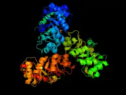
KDPG Aldolase was recently determined to be a trimer through crystallographic three-fold symmetry, with 225 residues.[2][3] The enzyme was determined to have a molecular weight of 23,942.[4] The trimer is stabilized primarily through hydrophobic interactions. The molecule has tertiary folding similar to triosephosphate isomerase and the A-domain of pyruvate kinase, forming an eight strand α/β-barrel structure.[3][5] The α/β-barrel structure is capped on one side by the N-terminal helix. The other side, the carboxylic side, contains the active site.[6] Each subunit contains a phosphate-ion bound in position of the aldolase binding site.[7] It has been found that there are four cysteinyl groups present in each subunit, with two readily accessible and two buried in the subunit.[8]
The active site contains the zwitterionic pair Glu-45/Lys-133.[9] The Lysine, which is involved in the formation of the Schiff base is coordinated by a phosphate ion and two solvent water molecules.[6][7] The first water molecule serves as a shuttle between the Glutamate and the substrate, staying bound to the enzyme throughout the catalytic cycle.[7] The second water molecule is a product of the dehydration of the carbinolamine that leads to the formation of the Schiff base.[7] It also functions as the nucleophile during hydrolysis of the enzyme-product Schiff base, leading to the release of pyruvate.[7]
As of late 2007, 13 structures have been solved for this class of enzymes, with PDB accession codes PDB: 1EUA, PDB: 1EUN, PDB: 1FQ0, PDB: 1FWR, PDB: 1KGA, PDB: 1MXS, PDB: 1WA3, PDB: 1WAU, PDB: 1WBH, PDB: 2C0A, PDB: 2NUW, PDB: 2NUX, and PDB: 2NUY.
Enzyme mechanism

One of the reactions KDPG Aldolase catalyzes, as in the Entner–Doudoroff pathway, is the reversible cleavage of 2-keto-3-deoxy-6-phosphogluconate (KDPG) into pyruvate and D-glyceraldehyde-3-phosphate.[9][10] This occurs through a stereospecific retro-aldol cleavage.[7] A proton transfer between the zwitterionic pair Glu-45/Lys-133 in the active site activates Lysine to serve as the nucleophile in the first step and Glutamate to aid in the base catalysis involved in the carbon-carbon cleavage.[9] Lysine Residue 133 serves as the nucleophile and attacks the carbonyl group of 2-Keto-3-deoxy-6-phosphogluconate to form a protonated carbinolamine intermediate, also known as a Schiff base intermediate.[7][9][10] The intermediate is stabilized by hydrogen bonding with residues in the active site.[9] A three carbon residue, glyceraldehyde 3-phosphate, is cleaved off through base catalysis with a water molecule and residue Glu-45.[7][9] The pyruvate is generated through the nucleophilic attack of water on the Schiff-base to reform a ketone. Aromatic interaction with Phe-135 ensures the stereospecific addition involved in the reverse process.[9]
KDPG aldolase has also been shown to catalyze the exchange of hydrogen atoms of the methyl groups of pyruvate with protons of the solvent.[10]

Evolutionary significance
History
Arguments have been made for both the convergent and divergent evolution of α/β-barrel structured enzymes such as KDPG Aldolase, triosephosphate isomerase, and the A-domain of pyruvate kinase.
Convergent evolution can lead to geometrically similar active sites while each enzyme has a distinct backbone conformation. Convergence to a common backbone structure, as is the case here however, has not been observed, although it is argues that it might be possible for a symmetrically repetitive structure as the one observed here.[11] The similarity in the folding of the three enzymes and the exceptional symmetry commonly suggests divergent evolution from a common ancestor. The functional similarity of the enzymes remains the strongest argument for divergent evolution.[11] All three enzymes activate a C–H bond adjacent to a carbonyl group. The active sites are located at the carboxylic ends of the β strands. Such congruence is in favor of divergent evolution.
Should the divergent evolution hypothesis prevail, this would suggest the existence of a class of enzymes with unrelated amino acid sequences yet analogous symmetrical structure and folding.[11]
Directed Evolution
KDPG aldolase has limited utility due to its high specificity for its natural substrates in the cleavage of KDPG and the reverse addition of D-glyceraldehyde-3-phosphate and pyruvate.[12] In vitro evolution has allowed KDPG aldolase to be converted into a more efficient aldolase with altered substrate specificity and stereoselectivity thereby improving its utility in asymmetric synthesis.[13] Rather than modifying the recognition site, the substrate is modified by moving the active site lysine from one β strand to a neighboring one.[13][14] The evolved aldolase is capable of accepting both D- and L-glyceraldehyde in their non-phosphorylated form.[15]
References
- ↑ Peekhaus N, Conway T (July 1998). "What's for dinner?: Entner–Doudoroff metabolism in Escherichia coli". J. Bacteriol. 180 (14): 3495–502. doi:10.1128/JB.180.14.3495-3502.1998. PMC 107313. PMID 9657988.
- 1 2 Mavridis IM, Tulinsky A (October 1976). "The folding and quaternary structure of trimeric 2-keto-3-deoxy-6-phosphogluconic aldolase at 3.5-A resolution". Biochemistry. 15 (20): 4410–7. doi:10.1021/bi00665a010. PMID 974067.
- 1 2 Mavridis IM, Hatada MH, Tulinsky A, Lebioda L (December 1982). "Structure of 2-keto-3-deoxy-6-phosphogluconate aldolase at 2 . 8 A resolution". J. Mol. Biol. 162 (2): 419–44. doi:10.1016/0022-2836(82)90536-8. PMID 7161801.
- ↑ Suzuki N, Wood WA (April 1980). "Complete primary structure of 2-keto-3-deoxy-6-phosphogluconate aldolase". J. Biol. Chem. 255 (8): 3427–35. doi:10.1016/S0021-9258(19)85716-2. PMID 6988426.
- ↑ Richardson JS (September 1979). "The singly-wound parallel beta barrel: a proposed structure for 2-keto-3-deoxy-6-phosphogluconate aldolase". Biochem. Biophys. Res. Commun. 90 (1): 285–90. Bibcode:1979BBRC...90..285R. doi:10.1016/0006-291X(79)91622-X. PMID 496979.
- 1 2 Bell BJ, Watanabe L, Rios-Steiner JL, Tulinsky A, Lebioda L, Arni RK (August 2003). "Structure of 2-keto-3-deoxy-6-phosphogluconate (KDPG) aldolase from Pseudomonas putida". Acta Crystallogr. D. 59 (Pt 8): 1454–8. doi:10.1107/S0907444903013192. PMID 12876349.
- 1 2 3 4 5 6 7 8 Fullerton SW, Griffiths JS, Merkel AB, et al. (May 2006). "Mechanism of the Class I KDPG aldolase". Bioorg. Med. Chem. 14 (9): 3002–10. doi:10.1016/j.bmc.2005.12.022. PMC 3315828. PMID 16403639.
- ↑ Möhler H, Decker K, Wood WA (July 1972). "Structure of 2-keto-3-deoxy-6-phosphogluconate aldolase. IV. Structural features revealed by treatment with urea and Ellman's reagent". Arch. Biochem. Biophys. 151 (1): 251–60. doi:10.1016/0003-9861(72)90495-x. PMID 5044518.
- 1 2 3 4 5 6 7 Allard J, Grochulski P, Sygusch J (March 2001). "Covalent intermediate trapped in 2-keto-3-deoxy-6- phosphogluconate (KDPG) aldolase structure at 1.95-A resolution". Proc. Natl. Acad. Sci. U.S.A. 98 (7): 3679–84. Bibcode:2001PNAS...98.3679A. doi:10.1073/pnas.071380898. PMC 31111. PMID 11274385.
- 1 2 3 Meloche HP, Glusker JP (July 1973). "Aldolase catalysis: single base-mediated proton activation". Science. 181 (4097): 350–2. Bibcode:1973Sci...181..350M. doi:10.1126/science.181.4097.350. PMID 4719907. S2CID 37940739.
- 1 2 3 Lebioda L, Hatada MH, Tulinsky A, Mavridis IM (December 1982). "Comparison of the folding of 2-keto-3-deoxy-6-phosphogluconate aldolase, triosephosphate isomerase and pyruvate kinase. Implications in molecular evolution". J. Mol. Biol. 162 (2): 445–58. doi:10.1016/0022-2836(82)90537-X. PMID 7161802.
- ↑ Farinas ET, Bulter T, Arnold FH (December 2001). "Directed enzyme evolution". Curr. Opin. Biotechnol. 12 (6): 545–51. doi:10.1016/S0958-1669(01)00261-0. PMID 11849936.
- 1 2 Fong S, Machajewski TD, Mak CC, Wong C (November 2000). "Directed evolution of D-2-keto-3-deoxy-6-phosphogluconate aldolase to new variants for the efficient synthesis of D- and L-sugars". Chem. Biol. 7 (11): 873–83. doi:10.1016/S1074-5521(00)00035-1. PMID 11094340.
- ↑ Wymer N, Buchanan LV, Henderson D, et al. (January 2001). "Directed evolution of a new catalytic site in 2-keto-3-deoxy-6-phosphogluconate aldolase from Escherichia coli". Structure. 9 (1): 1–9. doi:10.1016/S0969-2126(00)00555-4. PMID 11342129.
- ↑ Huisman GW, Gray D (August 2002). "Towards novel processes for the fine-chemical and pharmaceutical industries". Curr. Opin. Biotechnol. 13 (4): 352–8. doi:10.1016/S0958-1669(02)00335-X. PMID 12323358.
Further reading
- Meloche HP, Wood WA (1964). "Crystallization and characteristics of 2-keto-3-deoxy-6-phosphogluconic aldolase". J. Biol. Chem. 239 (10): 3515–3518. doi:10.1016/S0021-9258(18)97753-7. PMID 14245411.
External links
 Media related to 2-dehydro-3-deoxy-phosphogluconate aldolase at Wikimedia Commons